

Mole Removal - Can I Just Laser It Off?

Updated: Jul 17
In the clinic, I often meet patients who ask if a mole can “just be lasered off.” It sounds simple - quick, clean, no downtime. But when it comes to moles, the right approach depends on what the lesion truly is. Sometimes, it’s not just about removing what’s visible - it’s about knowing what lies beneath.
Case 1 - The Mole That Might Not Be a Mole
A GP referred a patient for a dark lesion on the left ear, concerned it might be melanoma. The patient had Fitzpatrick IV skin, which made photo triage unreliable - the lesion blended with surrounding pigment and texture.
Because of the GP’s concern, I recommended in-person evaluation and, if indicated, same-day excision with local flap reconstruction under local anaesthesia - allowing complete removal, immediate repair, and histology. The patient chose the subsidised referral via the polyclinic pathway, which is reasonable (for medically indicated lesions managed privately, Medisave and/or insurance can often offset costs). Fortunately, following excision biopsy, the histology returned as benign and not cancerous.
When melanoma cannot be excluded, laser is inappropriate - it destroys diagnostic tissue and affects future management plans.
Case 2 - When Laser Is the Right Tool
A Fitzpatrick II–III female came after multiple laser sessions elsewhere for flat “moles” on the forearms that only lightened slightly. She also had two slightly raised peri-oral moles (one on the philtrum, one next to the lower lip) and asked if these could be lasered too.
We started afresh: history, physical examination, and documentation to remove diagnostic uncertainty. Clinical assessment was in keeping with benign naevi, so I proceeded with CO₂ laser excision under topical anaesthesia:
Philtrum mole: treated to allow cosmetically sensitive secondary intention healing in the natural concavity.
Lower lip mole: lasered off entirely, with acceptance that a trace of pigment might remain. In this case, because the mole was slightly bigger, I decided to put in a couple of fine stitches to close the wound for a better cosmetic result.
Forearm flat moles: a focused repeat CO₂ session, with counselling on post-inflammatory hyperpigmentation (PIH) risk given her skin type.

This illustrates that laser can be appropriate - when the diagnosis is clear, expectations are aligned, and technique is tailored.
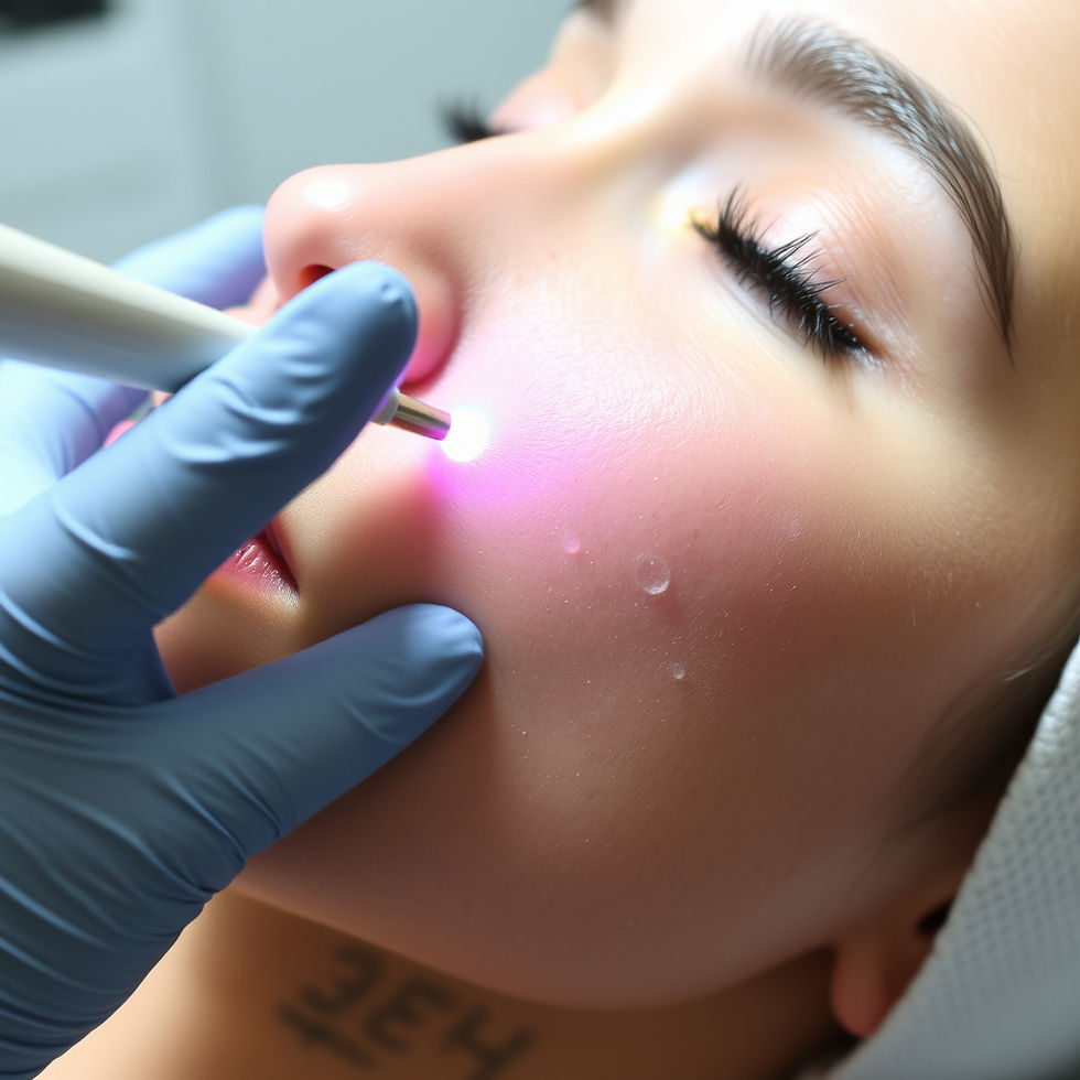
Case 3 - Raised Mole with Reported Changes: When Surgery Is the Only Appropriate Answer
A patient presented with a raised mole of about 1 cm size at the right temple, approximately one centimetre above the eyebrow. He reported that the lesion had changed in size over time.
Two features made laser immediately inappropriate here. First, the morphology: raised moles have deeper components that laser cannot reliably address in full. Surface ablation of a raised mole risks incomplete removal, regrowth, and most critically, destruction of the tissue needed for histological assessment in the laboratory. Second, the reported change in size. Any mole with a history of change warrants excision and histology, not surface treatment. Laser forecloses the diagnosis; so if it was cancer, it would have been missed.
The lesion was excised under local anaesthesia as a same-day procedure. The location of the mole required additional care: the frontal branch of the facial nerve (which enables one to raise the eyebrows) runs in this vicinity, and preservation of nerve function is a consideration that goes beyond simple mole removal. Excision and wound closure was performed with this anatomy in mind.
Histology returned and confirmed a benign naevus. The patient now has diagnostic certainty (and reassurance), a well-healed wound, and no residual lesion.
Case 4 - When a "Mole" Is Not a Mole: The Preauricular Sinus
A patient presented with two lesions around the ear - a raised mole in front of the earlobe, and a flat lesion in front of the tragus that she assumed was also a mole.
The raised lesion in front of the earlobe was straightforward to assess and plan for. The flat lesion in front of the tragus was not a mole at all. On examination, it was consistent with a preauricular sinus, a small congenital pit or tract that sits just in front of the tragus, representing a remnant of embryonic ear development. It is not uncommon, but it is easily misidentified as a skin lesion by patients, and occasionally by doctors who are not looking for it.
The distinction matters enormously. Lasering a preauricular sinus would not treat it as the sinus tract extends beneath the skin surface and laser addresses only what is visible. Worse, scarring over the sinus opening could obstruct drainage, leading to recurrent infection, abscess formation, and a significantly more complex surgical problem than the original presentation. The correct management of a preauricular sinus is surgical excision of the entire tract, not surface treatment of any kind.
This case illustrates a principle that runs through every case in this post: the treatment follows the diagnosis. A flat lesion in front of the tragus that has never been properly examined cannot be assumed to be a mole until proven otherwise.
Case 5 - When a "Routine" Mole in a Young Patient Is Anything But
A 17-year-old Caucasian patient was brought in by her mother for assessment of what appeared to be an innocuous mole on the cheek. They were understandably anxious about scarring as her younger sister had undergone surgical excision elsewhere and was left with rail-track marks from the sutures. Hence, they sought out a plastic surgeon this time, and specifically one with experience treating Caucasian skin.
As simple as it may seem, I know never to underestimate such moles and took a detailed history. What emerged was a constellation of risk factors that changed the entire assessment. She had a history of sunburns from a young age with no routine sun protection. Her family history included multiple members with basal cell carcinoma. And most significantly, the mole had already been lasered off by an aesthetic doctor locally, without any inquiry into her medical history. It had since recurred.
Given the size of the lesion, its location, the history of recurrence after laser, and the clinical risk profile, I opted for an incision biopsy using a 5mm punch under local anaesthesia rather than an immediate wide excision. My reasoning was: if histology returned something more sinister, the excision margins would need to be wider anyway. Therefore, establishing the diagnosis first avoids unnecessary tissue removal in a young patient, especially in a cosmetically sensitive area on the face.
Histology returned as a compound melanocytic naevus with an element of dysplasia (basically cells starting to turn abnormal). Not melanoma, but not entirely benign either. She will need regular mole surveillance, as she carries an elevated overall risk of melanoma.
Now consider what the previous treatment represented. The mole was lasered without any history being taken. If that laser had completely destroyed the lesion, and if the lesion had been a melanoma rather than a dysplastic naevus, there would have been no tissue for diagnosis, no further treatment, and no follow-up. Melanoma in young Caucasians is not a theoretical risk. It happens, and I have seen and treated them during my time in the UK and Europe.
An undiagnosed melanoma treated with laser is not a treated melanoma. It is a missed melanoma as surgical margins are required to reduce their risk of recurrence (not visible to the naked eye). It is essentially Russian rolette, a gamble with consequences that cannot be undone. Pause and contemplate that.
Why Histology and Context Matter
You cannot always reliably separate benign from malignant lesions by appearance alone, especially across skin tones
Not every skin lesion is what it appears. For example, a preauricular sinus presenting as a flat pit in front of the tragus can easily be mistaken.
Correct identification before any treatment is the only safe starting point.
Histopathology provides diagnostic certainty when there’s doubt and guides follow-up and further management (if indicated)
Method selection (laser, shave, surgical excision) should consider depth, site, skin type, scar tolerance, and diagnostic needs - not convenience
Takeaway
“Just lasering it off” may sound convenient - but what’s quick isn’t always safe or appropriate. The best results come from clear diagnosis first, then a method matched to lesion biology, location, skin type, and patient priorities.
If you’re considering mole removal, ask:
Will tissue be sent for histology if needed?
Is this complete removal or surface-only?
How will scarring, post-inflammatory hyperpigmentation (PIH) risk, and follow-up be managed?
For moles requiring surgical excision and histological assessment, this can be arranged as a same-day procedure.
Written by Dr Ng Zhi Yang, UK & Singapore accredited Plastic Surgeon.


Comments