

Ultrasound Evaluation before Revision Rhinoplasty

Updated: Sep 27
Revision rhinoplasty is almost certainly never a simple repeat of the first operation. By the time a patient is considering a second or third procedure, the anatomy has usually been changed, sometimes by surgery, sometimes by scarring, often by both. Working out exactly what's underneath the skin, rather than assuming, is often the difference between a plan that works and one that doesn't.
Point-of-care ultrasound (POCUS) has become a routine part of my practice. The cases below show how and why in patients considering revision rhinoplasty.
Case 1: The Dent That Two Surgeries Didn't Explain
A patient came to me with concerns about a "dent" around the scroll area of her nose. Her history was complicated. A nasal bone fracture had for some reason, gone uncorrected during an initial rhinoplasty that focused on the tip instead. Significant scarring followed, treated with multiple rounds of steroid injections without success. A revision abroad then used what sounded like a cadaveric rib allograft. Three years later, she presented with a "dent" that had not resolved with steroid injections. Attempts to camouflage with HA filler were less than satisfactory and she has had them dissolved.
The literature suggests resorption rates as high as 30 percent in irradiated rib allografts, but in this patient it was academic: a POCUS showed no sonographic evidence of any remaining graft material at all. The scan also mapped the bony architecture directly, confirming a clear step-off deformity at the old fracture site, and measured the soft tissue thickness at 5.7mm (see picture below).

That combination of findings, no graft, a healed-in-the-wrong-position fracture, and a specific tissue thickness, meant I could rule out further steroid injection (the upper lateral cartilages were too close for that to be safe) and instead design a targeted plan: cartilage grafting for the bony depression, combined with supratip scar excision and tip refinement. Without the scan, the working assumption might reasonably have been "the graft is still there, just resorbing unevenly." That assumption would have led to the wrong plan.
Case 2: Seven Rhinoplasties, and a Plan I Almost Started Without Scanning
Another patient had been through seven prior rhinoplasties and came in with significant fullness and stiffness across the nose. My initial instinct, based on the visible pores and what looked like thick, oily skin, was to treat this with oral retinoids and CO2 laser resurfacing.
POCUS changed that plan before it started. A longitudinal scan showed a dense, hyperechoic band of mature fibrosis running along the nose (see picture below). The transverse view confirmed soft tissue thickening throughout, most pronounced in the supratip region, at roughly 5 mm, essentially replaced by scar tissue rather than oily skin. This is a pattern I've written about in my own published research on tip control in closed rhinoplasty: inadequate closure of dead space after multiple procedures tends to show up exactly here.

That single finding changed the entire approach. Instead of resurfacing skin that wasn't actually the problem, I moved to intralesional triamcinolone injections at a higher concentration than usual, given how dense the scar was, placed across the thickest areas as confirmed by the scan and by the patient's own sense of where things felt tightest. He's now being monitored at six-week intervals to allow the steroid to work on collagen remodelling (see below).

I recently saw him for a third time and POCUS assessment showed that the scar tissue had reduced to about 3.3 mm (from approximately 5 mm initially) (see above) and he was delighted with his overall results, especially given the multiple surgeries he had gone through previously.
Case 3: A High Radix After Implant Removal, and Why I Didn't Go Straight Back to Surgery
A third patient travelled from Malaysia, where he's currently based, to see me. He has had a silicone implant placed in his nose over ten years ago, then had it removed the year before in Cambodia. The surgeon there had placed a dermofat graft, confirmed by a donor site scar in the groin crease, in its place. His complaint was specific - the radix was too high, and he wanted it flatter.
Going by history alone, the natural assumption is that a year-plus-old dermofat graft would have settled into a reasonable amount of bulk. To lower the radix now would mean surgery to excise or revise the graft. POCUS however, showed something different: only faint scar tissue around the radix, far less substance than I expected (see below).

The finding that the dermofat graft has likely resorbed, is not totally unexpected as resorption rates have been reported between 18-45%, and higher in the radix. This changed the sequence of treatment rather than the eventual goal. Instead of going straight back to surgery, we agreed to try steroid injections first, to see how much reduction we could achieve non-surgically before committing to another procedure in an area that's already been operated on twice as further surgery may lead to further scarring, which would not only fail to achieve the patient's desired outcome, but may potentially make it worse through thickened scars. This patient subsequently went on to have surgery as his condition did not improve with the steroid injection.
Case 4: The High Radix & The Pinched Tip
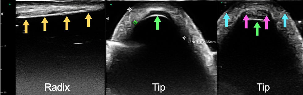
A patient consulted for a second opinion for a revision rhinoplasty. Her history revealed a primary silicone augmentation which then developed extrusion about two years later, removal and re-implantation with another silicone implant just three months after. She came in bothered by a "pinched" tip, where ear cartilage grafts were placed, along with a dorsum that was not as straight as she had hoped for.
She did not notice the prominent, high-riding radix on lateral views. I explained to her that that was the most obvious finding, to me, actually. Interestingly, she then said that a third surgeon whom she had enquired with, and would go to consult next, mentioned the same. In my mind, there were a few possibilities here - swelling, scarring and an overprojected tail of the silicone implant (which I thought was most likely based on examination). As for the tip, it could again be one of these, or a mixture, which could then contribute towards the less-than-straight profile which she desired.
Once again, POCUS enabled objective assessment immediately. It visualised the hypoechoic profile of the silicone tail extending beyond the nasal bone to the nasofrontal angle beneath a thin skin envelope. When compared against the POCUS scan of pure post-removal fibrous scar tissue in Case 3 above (which was a dense, hyperechoic white footprint), the mechanical cause was unquestionable. For the "pinched" tip, the POCUS views suggest that the hypoechoic silicone implant was now positioned posteriorly, possibly due to migration, or it may also be sandwiched between the medial crura and ear cartilage grafts (see below).
What These Cases Have in Common
In the cases above, the visible problem (a dent, a stiff and oily-looking nose, a radix that sat too high) had an assumed cause that turned out to be incorrect, and in each case the wrong assumption came from surgeons, including at points, me, working from examination or clinical history alone. POCUS didn't replace clinical judgment, but it gave me something clinical examination alone couldn't - an objective answer to "what is actually there," before committing to a plan.
For patients considering revision rhinoplasty in particular, that matters far more than in primary cases. Anatomy that's been operated on multiple times rarely behaves the way an unoperated nose does, and the cost of guessing wrong is another procedure on tissue that has less room for error each time.
Written by Dr Ng Zhi Yang, UK & Singapore accredited Plastic Surgeon.
References
Rohrich RJ, Dayan E, Durand PD, Brito I, Gronet E. Warping Characteristics of Rib Allograft Cartilage. Plast Reconstr Surg. 2020 Jul;146(1):37e-42e. doi: 10.1097/PRS.0000000000006896. PMID: 32590648.
Kosins AM, Obagi ZE. Managing the Difficult Soft Tissue Envelope in Facial and Rhinoplasty Surgery. Aesthet Surg J. 2017 Feb;37(2):143-157. doi: 10.1093/asj/sjw160. Epub 2016 Dec 13. PMID: 27965218.
Ng ZY, Liu A, Shoaib T. Tip control in closed rhinoplasty. Eur J Plast Surg. 2026;49:98.
Aydın C, Yücel ÖT, Akçalar S, Atay G, Özer S, Sözen T, Akata D. Role of steroid injection for skin thickness and edema in rhinoplasty patients. Laryngoscope Investig Otolaryngol. 2021 Jul 9;6(4):628-633. doi: 10.1002/lio2.616. PMID: 34401482; PMCID: PMC8356887.
Kim JH, Ko HS, Park SW. Using Dermofat Grafting in Revision Rhinoplasty. Aesthetic Plast Surg. 2021 Apr;45(2):617-625. doi: 10.1007/s00266-020-01950-5. Epub 2020 Sep 30. PMID: 32997238.


Comments